|
Readers of this blog are already familiar with my lovely finds of Terataspis fragments, all found in the early spring (and I beg forgiveness for the false match from 2019 that turned out to be Echinolichas -- certainly a face palm moment!). But I can very confidently say that I am sitting on so many Terataspis fragments it is ridiculous. I've been spending an equally ridiculous time in the prep lab on these fragments, with nothing fully complete in terms of preparation. But I am one of the select few who has chunks of Terry sitting on his dining room table -- not that this is some benchmark of anything beyond being unbearably niche! Of course, they are set in some of the most brutal matrix ever devised by depositional powers. It is variable in hardness from soft sandy shale to the hardest crystalline quartz that requires very slow scribing and even the use of aluminum oxide. I have a few choice pieces entrusted to a seasoned preparator, but I still have tons of material on hand to get through on my own, the so-called B-pile frags. So, let's get to talking about Terry.
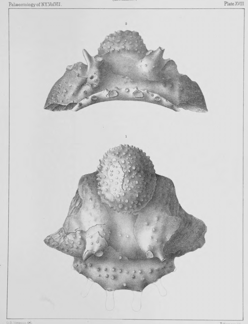
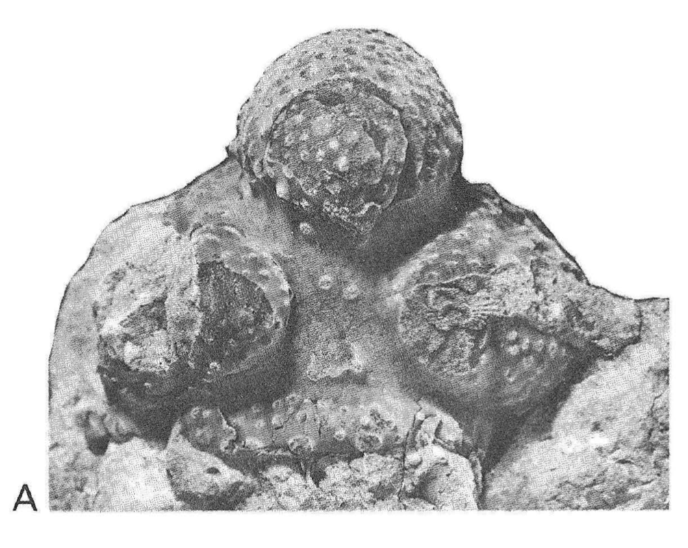
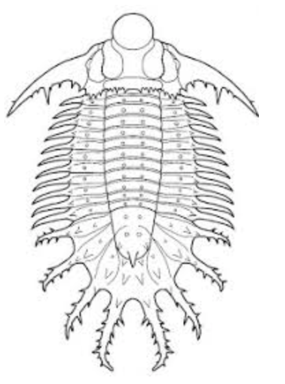
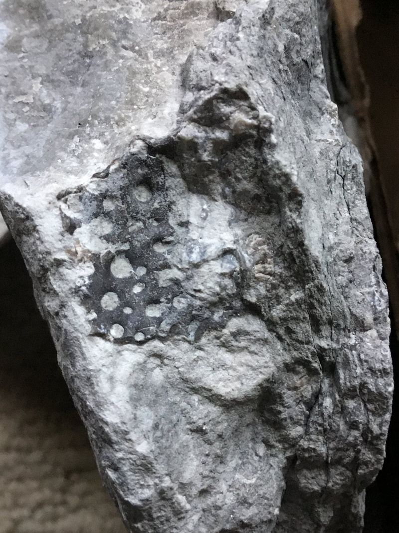
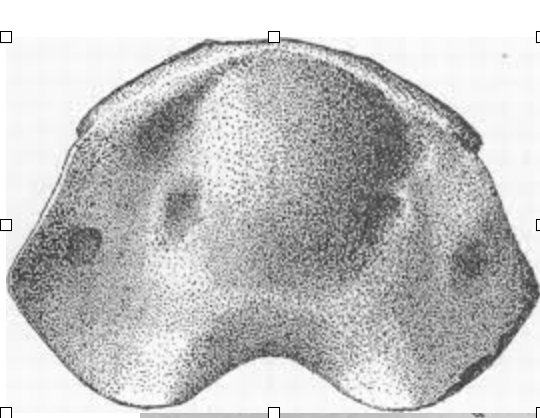
Here are some classic images of the cranidia, which includes the three glabellar lobes. All of the lobes have fine tubercles, but it is the median lobe that has the "moon rock" appearance. It is by far one of the more impressive and diagnostic features of this monotypic genus. The first image is a rendering in the classic Hall & Clarke plates,, and the second is a photograph of a specimen that was included in Ludvigsen's 1979 "green bible" of Ontario trilobites.
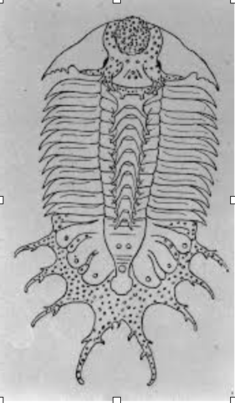
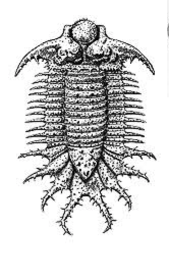
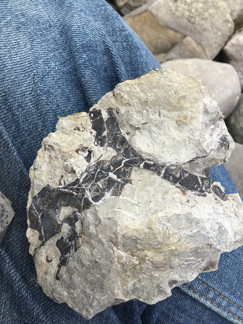
Terataspis has undergone some... cosmetic changes... over time on the basis of found fragments. It would not be until Riemann in 1941 that we more accurately approach the proper morphology of the trilobite. In the first incarnation of reconstruction, the cranidia and the pygidium is largely correct, but there was a mishap in attributing a large dalmanitid to account for being included for the pleurae, which doesn't quite lend it the right look. Only with more fragments found properly attributed could a more fulsome and accurate reconstruction come to the fore. I'm not even going to ask about the bizarre prong-spines along the axis! It should be noted that large organisms such as these will vary significantly in terms of their prosopon and other morphological features; some will have additional tubercles, spines, and even spinose protrusions not seen on other specimens. That is a feature, not a "bug" (ok, the pun was uncalled for!), just as any complex organisms will show different phenotypic variations (think of Homo sapiens and our rich diversity as an example of how we simply don't look identical). But let's move on to some of my own examples. 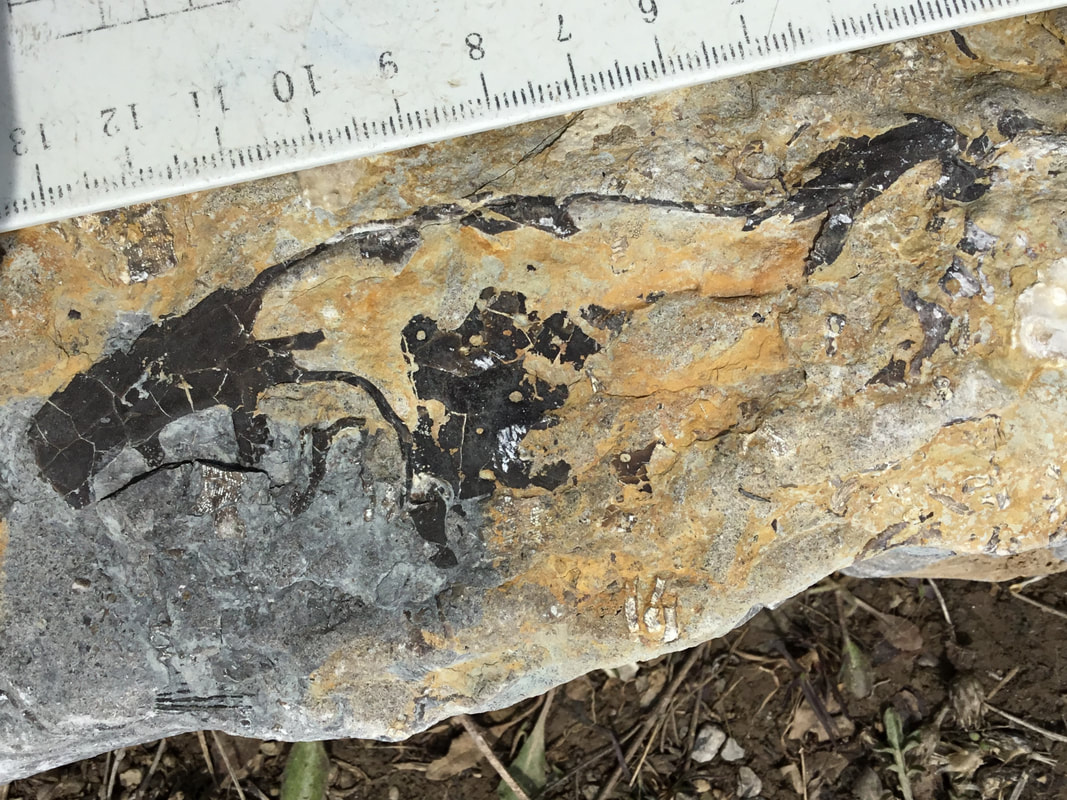 It certainly doesn't get more diagnostic than this. This is the left librigena, sadly missing some shell from having been exposed, and fairly flat in being preserved in a layer that is largely arenaceous and weathered. But already, the fragment is 13+ cm long, and it isn't even complete (complete cheek might be pushing over 15 cm -- that's shy of just half a foot!). The fractal-like spines appear here.
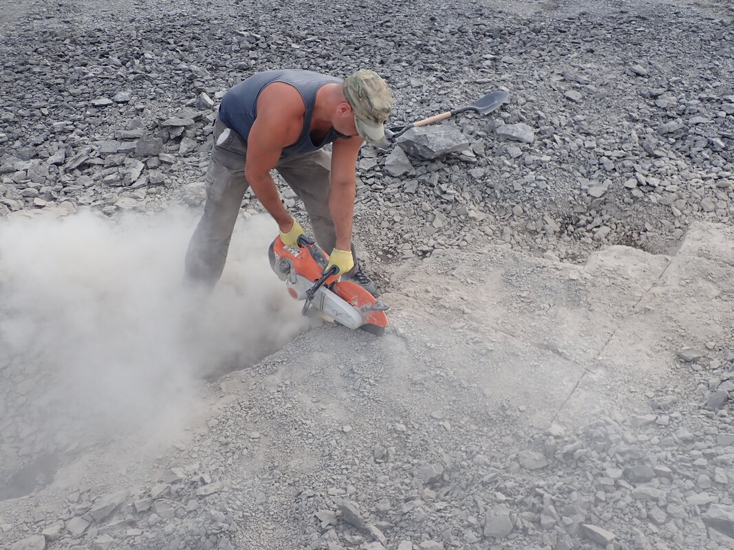
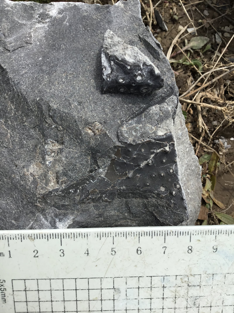
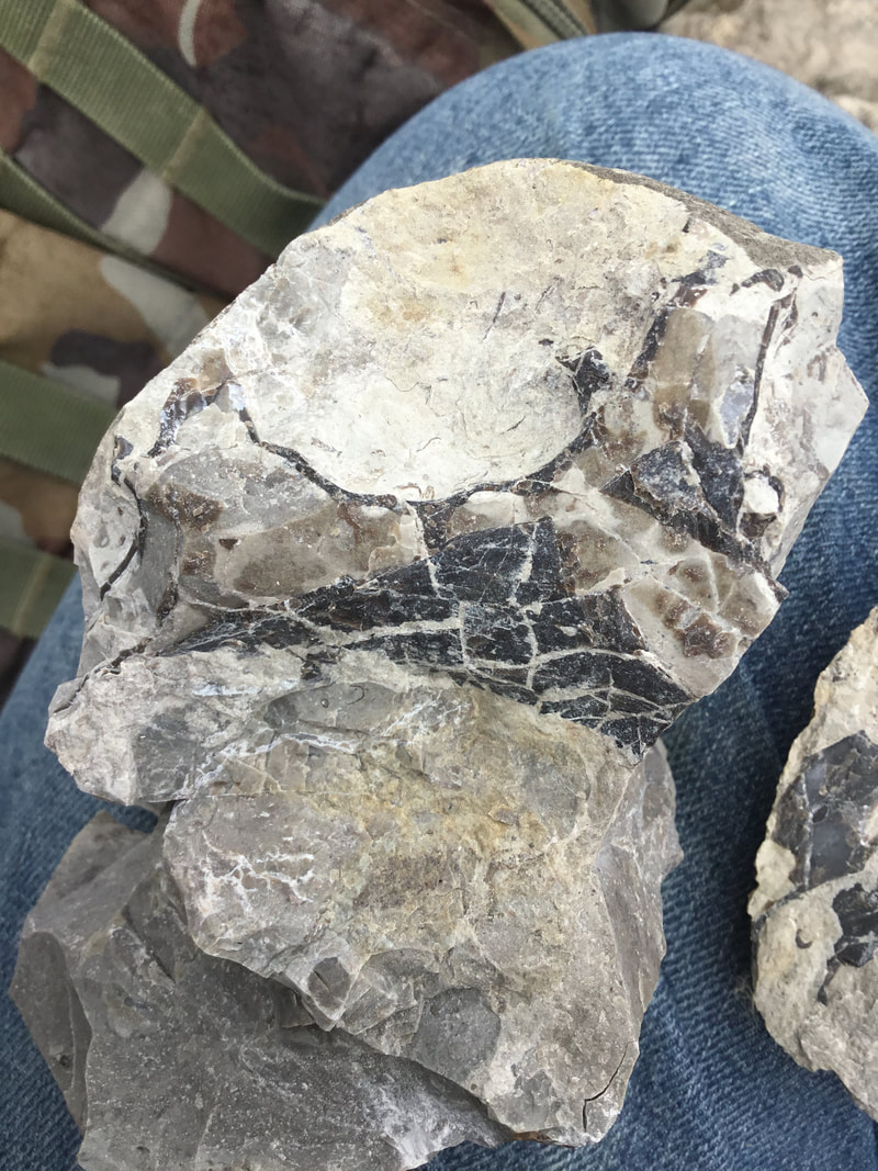
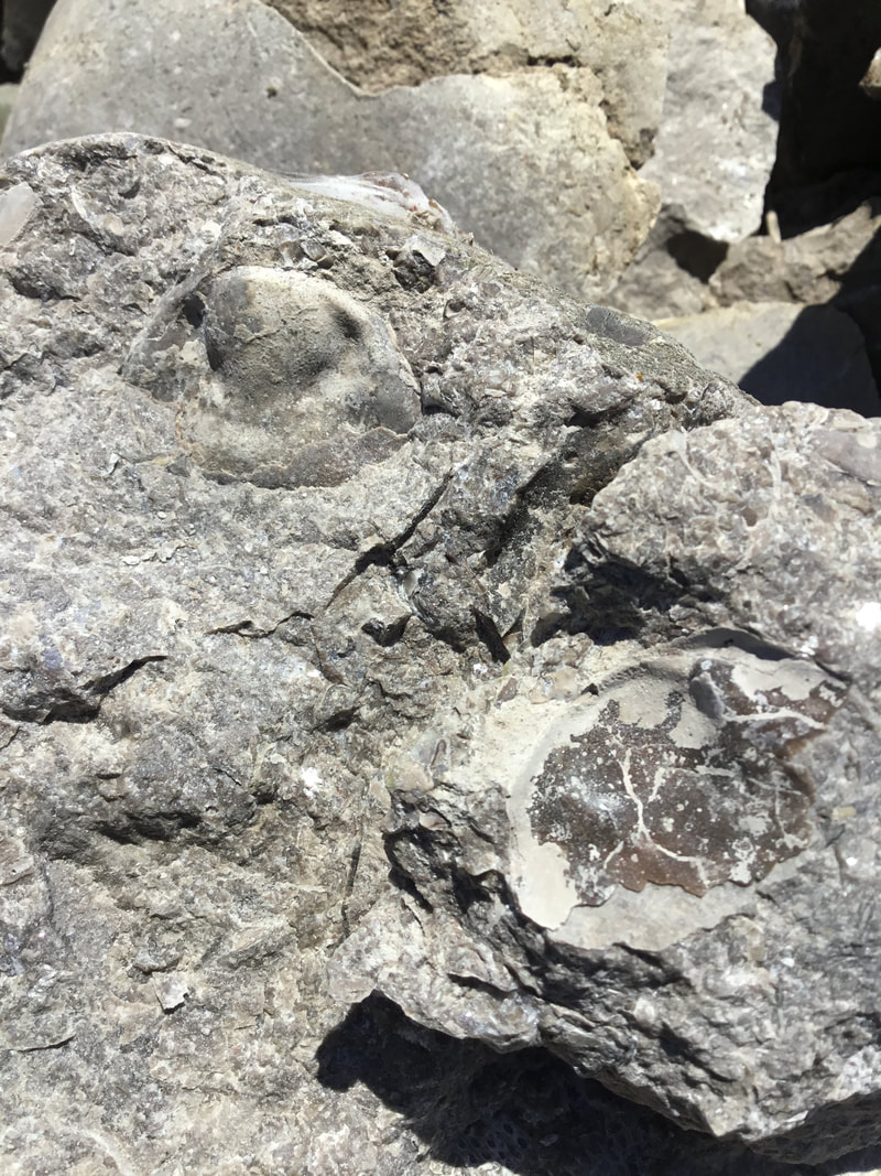
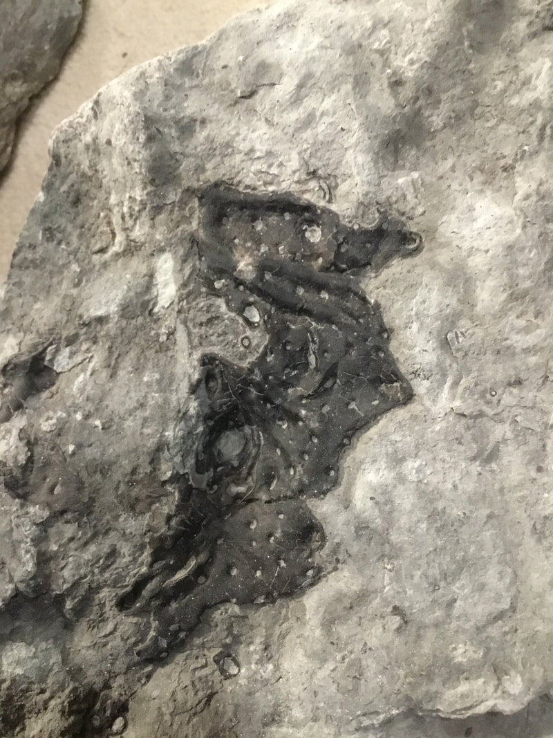
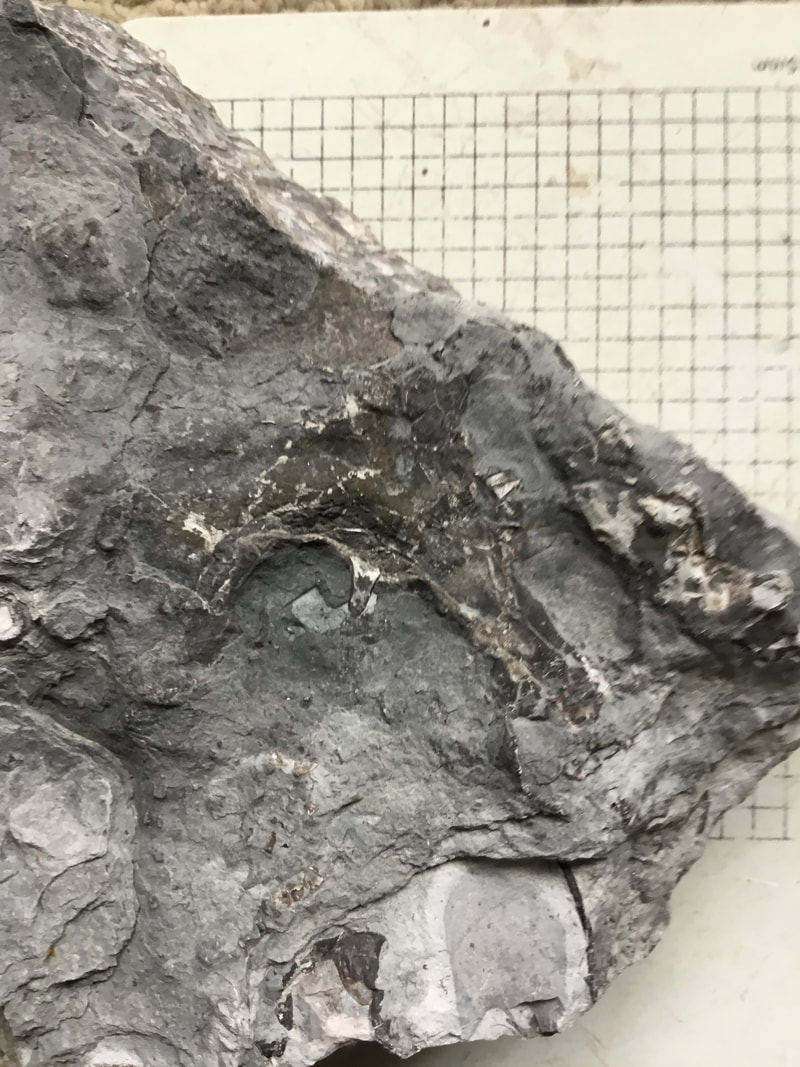
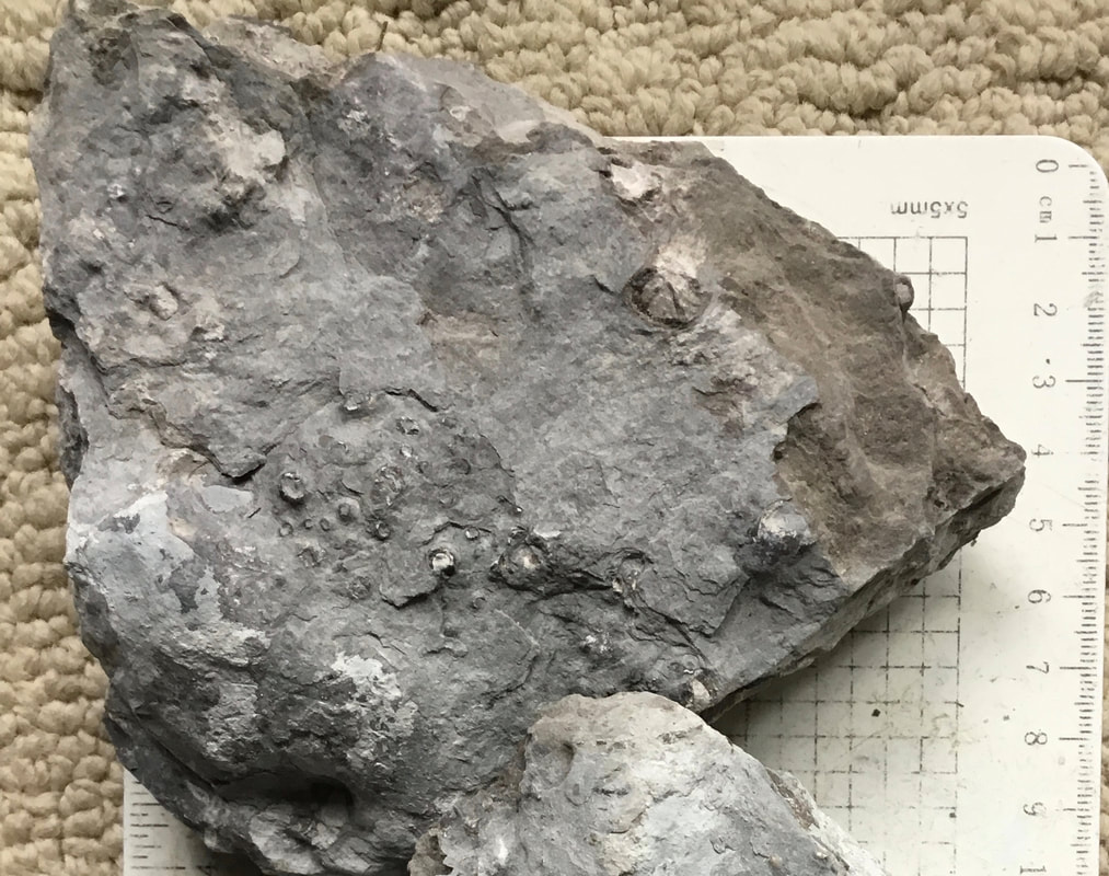
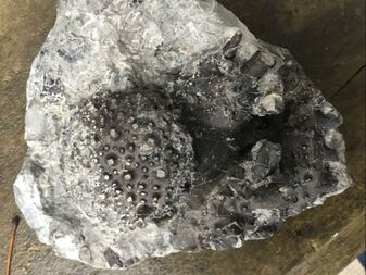
Gimme some skin! This material is "perversely tough" to quote Ludvigsen. You do not split this material because it is 70-90% chert by volume in Bois Blanc Formation, dominated by coral, riddled with crinoids and other fauna. These fragments occurred in the tough micritic horizon where one may be fortunate (as I have been) to find full prone Calymene platys, but the shell is rarely going to separate from the impression. But once you find telltale signs of Terataspis, you begin to find them everywhere. At my secret location, I was able to bucket a good number! Here are some in situ pictures of what the material in the field looks like. Very underwhelming! But keep in mind that this is only what is visible, with the rest encased in tough matrix. The inexperienced collector would likely not bother with these, thinking they are just tiny fragments. 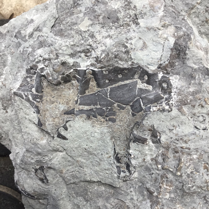 An example of what lurks beneath. I am far from done with this piece, but this is likely a piece of the pygidial spine, ventally oriented. 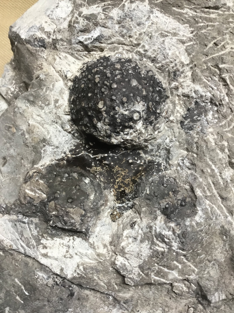 Another piece that might have been overlooked, I had already put a good innings on the cranidium of this specimen before handing it off to a seasoned preparator. I now have better tools and can manage my own workload. The left cheek will sadly not be there, but the right one might be... as well as the occipital ring... and who knows what else? The median glabella is about the size of a golfball. The initial field state only showed a few tubercles.
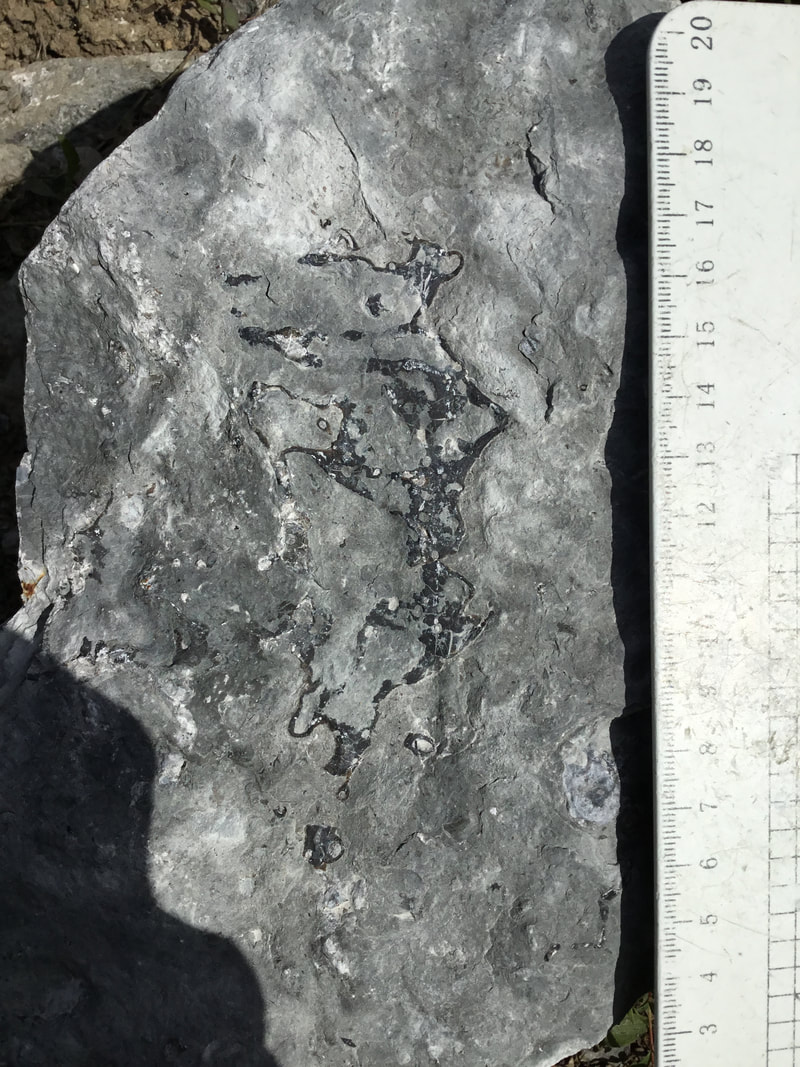
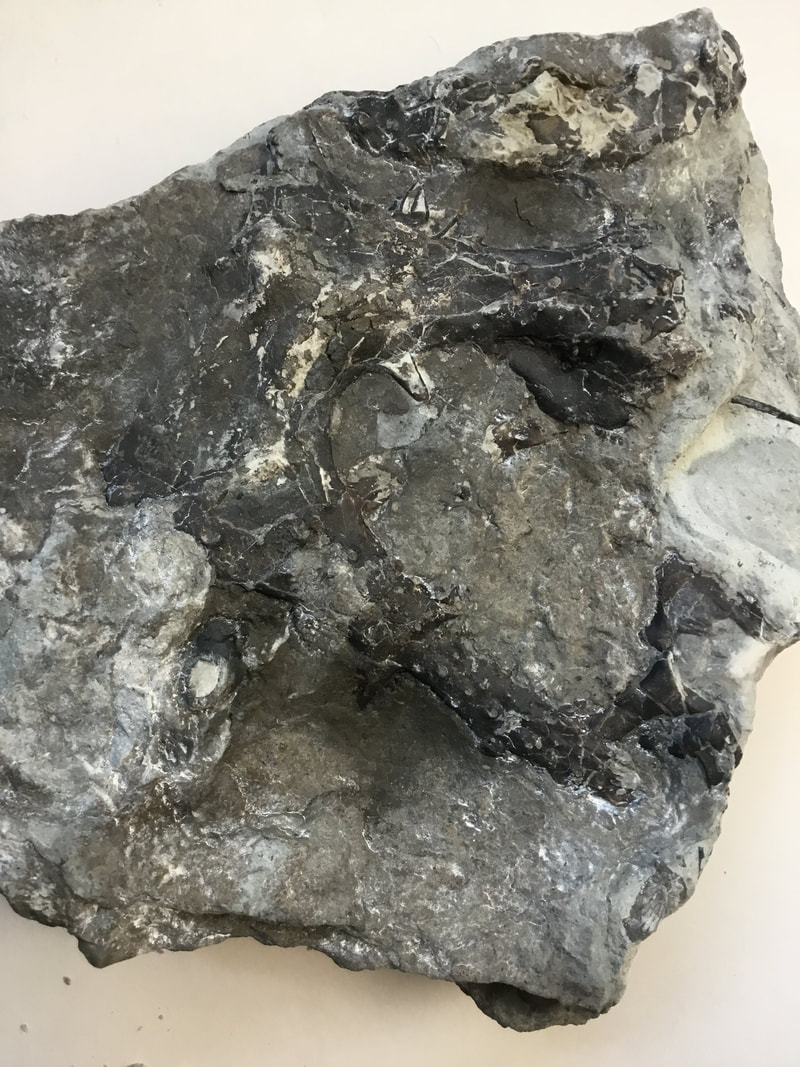
And even close to home! These two pieces are positive/negative of each other, part of four distinct pieces I found from glacial erratics here in London.
From the same glacial drift, a thumbprint-sized hypostome. Lichid hypostomes are not known for their fanciness compared to, say, Hypodicranotus. But here is a Terry hypostome I still need to prep.
From the field to the lab of my comrade, a ventral portion of the pygidium (sans spines). 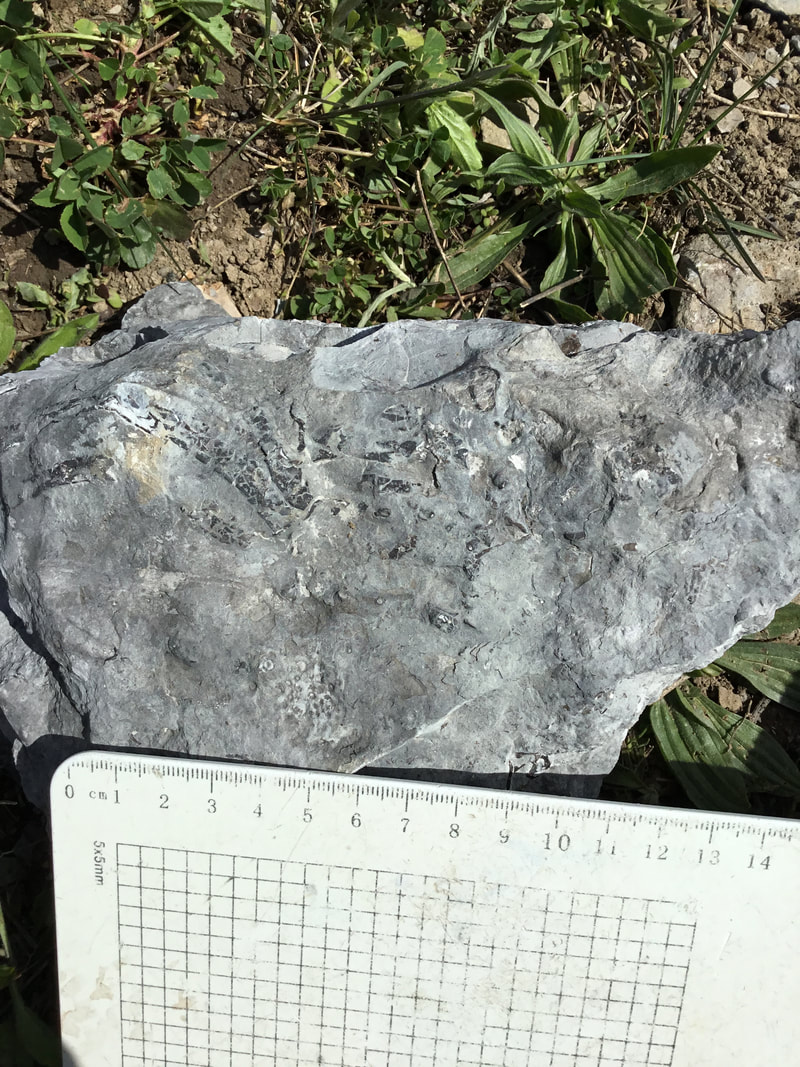 A chunk of thorax. I'm far from done with it, but it looks... crappy in the field. 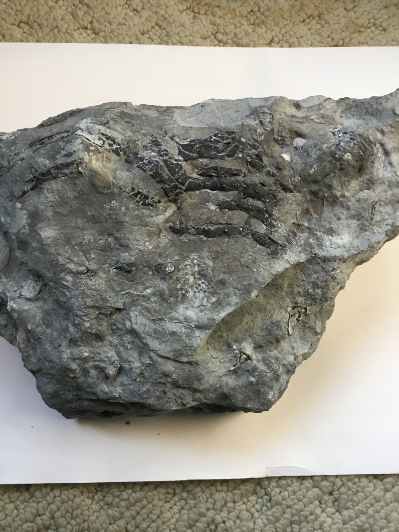 A bit of scribing and dolomite abrasion, and we're seeing a wee bit more. But if we look closely... 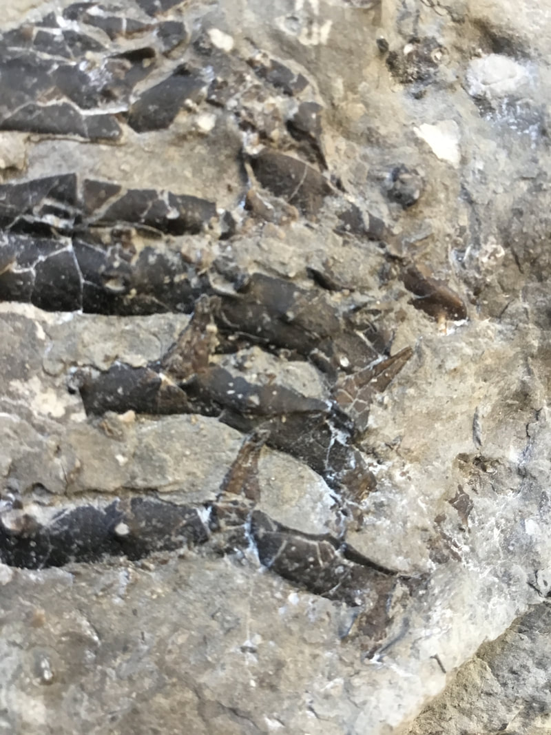 Spines... on the pleurae. These are not the end-of-pleurae spiny frills, but appearing before the tips of the pleurae. That variation I find exciting! 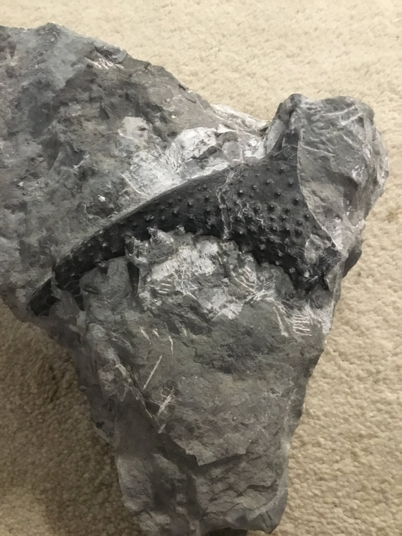 A left genal that is not yet completed by my master preparator and comrade. I have a few free cheeks lying around, moulty buggers!
Another work in progress. We have two apparent pygidial spines, but there is other specimens in the mix (see the occipital ring above, and there is a free cheek at the far right (I've since exposed that but not taken a photo yet). Moulting ground! (Or tidal sorting).
Like night and day. This chunk of cranidium took me 60+ hours, including going the nuclear option of using aluminum oxide on hard, crystalline chert that dolomite was too soft to touch. Those spinose tubers "fly" over the very deep furrow (4 cm) above the occipital groove. The median glabella already measures about two inches across and long. Sadly, it is crushed, and the anterior edge has a chert separation between the tubercles and the base. Not much more I can do with this piece, and sad other parts are missing. But here is a gallery of other shots, including texture (please pardon the dust!). These photos are rubbish since they do not convey the full depth and detail of having it in hand. There is more I could do with it, and I could spin a yarn of so much that was done to ensure it could get to this state. But this was great practice for the tons of material I still have left, with the possibility of some surprises. Obviously I have no photographed and shown every piece I've collected. I've just been picking at the smaller, easier stuff -- there are bigger specimens in the mix with some hope that they might be complete.
Comments are closed.
|
Kane Faucher
Archives
February 2024
|
 RSS Feed